Cystoid Macular Edema
What is cystoid macular edema (CME)?
Cystoid macular edema (CME) refers to swelling (edema) in the central part of the retina (macula) that occurs in the form of multiple fluid-filled spaces (cysts). It can occur in a variety of settings, but cataract surgery is the most common cause. Inflammation due to surgery sometimes makes the blood vessels within the central retina leaky. As a result, fluid leaks from these vessels into the retina, causing it to swell. Since the retina does not function properly when it is swollen, blurred vision results. In cases of CME following cataract surgery, the vision is often significantly improved shortly after surgery but becomes blurred within a few weeks as the retina becomes swollen.

How is CME diagnosed?
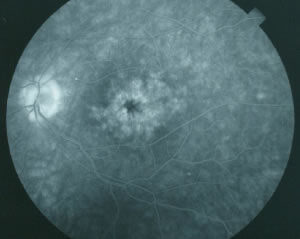
CME is sometimes visible during the eye exam. Its presence is usually confirmed using fluorescein angiography, a test that involves injecting a dye (fluorescein) into a vein in the arm and taking numerous photographs of the retina as the dye circulates through the blood vessels in the retina. If CME is present, the dye can be seen leaking into the retina. In some cases, the leakage is described as “petalloid”, because its appearance in the photographs resembles the petals of a flower. The image to the right is an example of a fluorescein angiogram that shows “petalloid” dye leakage in the central macula:
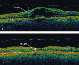
 CME can also be confirmed with optical coherence tomography (OCT), which shows the retina in cross-section. The image to the left shows how CME looks on OCT, before (above) and after (below) treatment:
How is CME treated?
Several treatments are available for CME. Below is a summary of the various options:
1) Eye drops are usually the first treatment used, since they involve minimal risk and are often effective. Two types of anti-inflammatory drops are typically used together. One is similar to aspirin in liquid form, and the other is similar to prednisone (a common “steroid” pill) in liquid form.
2) Periocular steroid injection is usually the second option, if drops have not improved the vision within several months of treatment. “Periocular steroid injection” means “injection of steroid medication around the eye”. This procedure is typically performed in the office with only topical anesthesia (i.e., numbing drops). It is virtually painless and takes just a few minutes.
3) Intraocular injections are often the third option. Triamcinolone, a steroid, is sometimes injected into the vitreous of the eye. Triamcinolone has two disadvantages: it can cause cataracts and glaucoma (elevated eye pressure). Avastin, a medicine often used for “wet” macular degeneration, can also be injected into the eye to reduce macular edema.
4) Vitrectomy surgery is the fourth option. “Vitrectomy” means “removal of the vitreous”. This procedure is performed in an operating room. The retina specialist places instruments inside the eye, through the sclera (the “white” of the eye), for removal of the vitreous, a clear gel in the back part of the eye.


